Book Description
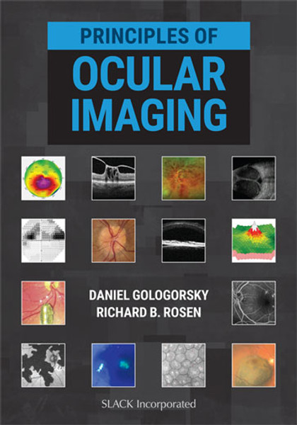
An essential text for the modern eye specialist, Principles of Ocular Imaging presents a comprehensive guide of all current ocular imaging modalities for ophthalmologists, optometrists, and those in training. Drs. Gologorsky and Rosen deliver a concise yet thorough overview of 22 imaging modalities unique to ophthalmology, emphasizing clinical application and replete with illustrative examples and ophthalmic images.
Principles of Ocular Imaging is divided into the following subspecialties for easy reference in busy clinical environments:
Principles of Ocular Imaging is divided into the following subspecialties for easy reference in busy clinical environments:
- Oculoplastics: external photography, ptosis visual fields, slit lamp photography, and orbital ultrasonography
- Cornea and refractive: corneal topography, confocal microscopy, anterior segment optical coherence tomography (AS-OCT), ultrasound biomicroscopy (UBM), biometry for intraocular lens (IOL) calculations
- Glaucoma: visual fields, optical coherence tomography (OCT) in glaucoma
- Retina: fundus photography, fluorescein angiography (FA), indocyanine green (ICG) angiography, fundus autofluorescence (FAF), OCT in retina, optical coherence tomography angiography (OCTA), adaptive optics (AO), microperimetry, retinal ultrasonography
- Neuro-Ophthalmology: electrophysiology of vision and computed tomography (CT) & magnetic resonance imaging (MRI)
More Information
Contents
CONTENTSDedication
About the Editors
Contributing Authors
Preface
Foreword
Introduction
Section I Oculoplastics
Section Editor: Wendy W. Lee, MD, MS
Chapter 1 External Photography
Alexandra E. Levitt, MD, MPH; Apostolos Anagnostopoulos, MD; and Wendy W. Lee, MD, MS
Chapter 2 Ptosis Visual Fields
Alexandra E. Levitt, MD, MPH; Apostolos Anagnostopoulos, MD; Ann Q. Tran, MD; and Wendy W. Lee, MD, MS
Chapter 3 Slit Lamp Photography
Ashwinee Ragam, MD
Chapter 4 Orbital Ultrasonography
Ying Chen, MD; Andrew J. Rong, MD; Amy Huang, BS; John Hinkle, MD; Nimesh Patel, MD; and Wendy W. Lee, MD, MS
Section II Cornea and Refractive
Section Editors: Ashwinee Ragam, MD and Oriel Spierer, MD
Chapter 5 Corneal Topography
Ashwinee Ragam, MD
Chapter 6 Confocal Microscopy
Ashwinee Ragam, MD
Chapter 7 Anterior Segment Optical Coherence Tomography
C. Maxwell Medert, MD; Hasenin Al-khersan, MD; and Ann Q. Tran, MD
Chapter 8 Ultrasound Biomicroscopy
Ashwinee Ragam, MD
Chapter 9 Biometry for Intraocular Lens Calculations
Ashwinee Ragam, MD
Section III Retina
Section Editors: Daniel Gologorsky, MD, MBA and Richard B. Rosen, MD
Chapter 10 Fundus Photography
Daniel Gologorsky, MD, MBA
Chapter 11 Fluorescein Angiography
Daniel Gologorsky, MD, MBA
Chapter 12 Indocyanine Green Angiography
Daniel Gologorsky, MD, MBA
Chapter 13 Fundus Autofluorescence
Hasenin Al-khersan, MD and Ann Q. Tran, MD
Chapter 14 Optical Coherence Tomography in Retina
Daniel Gologorsky, MD, MBA
Chapter 15 Optical Coherence Tomography Angiography
Chris Y. Wu, MD and Richard B. Rosen, MD
Chapter 16 Adaptive Optics
Chris Y. Wu, MD and Richard B. Rosen, MD
Chapter 17 Microperimetry
Hasenin Al-khersan, MD; Thomas Lazzarini, MD; and Ann Q. Tran, MD
Chapter 18 Retinal Ultrasonography
Daniel Gologorsky, MD, MBA and Yale Fisher, MD
Chapter 19 Electrophysiology of Vision
Alessandra Bertolucci, MD
Section IV Glaucoma
Section Editor: Stephen Moster, MD
Chapter 20 Visual Fields in Glaucoma
Stephen Moster, MD; Cindy X. Zheng, MD; and Michael M. Lin, MD
Chapter 21 Optical Coherence Tomography in Glaucoma
Michael M. Lin, MD; Cindy X. Zheng, MD; and Stephen Moster, MD
Section V Neuro-Ophthalmology
Section Editor: Wendy W. Lee, MD, MS
Chapter 22 Computed Tomography and Magnetic Resonance Imaging
Michelle W. Latting, MD; John W. Latting, MD; Sheikh Faheem, MD; and Wendy W. Lee, MD, MS
Bibliography
Financial Disclosures
Index
Reviews
"What a timely, practical, and informative textbook. A truly comprehensive resource on a multitude of ocular imaging modalities. A must-read for all health care professionals involved in vision care."—Eduardo C. Alfonso, MD, Professor and Chairman, Bascom Palmer Eye Institute, Kathleen and Stanley J. Glaser Chair in Ophthalmology
“Principles of Ocular Imaging is a beautiful, handy visual guide to all types of ocular imaging—from the traditional to the most cutting-edge. Gologorsky and Rosen have done an incredible job. A must-have for all ophthalmologists!”
—Amy Schefler, MD, Associate Professor of Clinical Ophthalmology, Weill Cornell Medicine, Houston Methodist Hospital and University of Texas Health Science Center of Houston
“This is an excellent textbook that provides insight about the various evolving imaging modalities for everyone from trainees to practicing ophthalmologists.”
—Ajay Kuriyan, MD, MS, Assistant Professor, Mid Atlantic Retina, Wills Eye Hospital, Thomas Jefferson University
“This beautifully illustrated 256-page text provides the reader with concise paragraphs of clinical wisdom followed by selected timely references. The topics range from Oculoplastics to Neuro-Ophthalmology and Glaucoma. My favorite sections were Retina and Cornea, which showcase the latest technologies. Easy reading and excellent images make this book useful for all clinicians.”
—Harry W. Flynn, MD, Professor of Ophthalmology, J. Donald M. Gass Chair in Ophthalmology, Bascom Palmer Eye Institute“The emergence of ocular imaging technologies over the last 2 decades has changed the landscape of eye care. No longer are advanced technologies only found in specialty practices or academic university settings, but instead are being utilized in all kinds of practices across the country by both optometrists and ophthalmologists—ultimately allowing us to take better care of our patients.
“The textbook Principles of Ocular Imaging by Daniel Gologorsky, MD, and Richard Rosen, MD is a comprehensive guide to 22 imaging technologies that are widely used in eye care. It not only includes a detailed review of each imaging technology but provides the most clinically relevant aspects as well as great examples highlighting each of the technologies. This will become one of those must-have textbooks for every practice and will become the foundation for understanding imaging in eye care.”
—Mark T. Dunbar, OD, FAAO, Director of Optometric Services, Bascom Palmer Eye Institute
About the Editors
Daniel Gologorsky, MD, MBA is an ophthalmologist and vitreoretinal specialist trained at the Bascom Palmer Eye Institute and the New York Eye and Ear Infirmary. He completed his undergraduate studies at Cornell University, and obtained his MD and MBA degrees from Dartmouth Medical School and the Tuck School of Business at Dartmouth, respectively. He has authored more than 50 peer-reviewed publications and textbook chapters, and has lectured extensively at national and international ophthalmological conferences. Dr. Gologorsky serves as the Chief of Ophthalmology at Broward Health Medical Center in Fort Lauderdale, Florida. Dr. Gologorsky enjoys teaching and is an avid history aficionado, with special interests in classical Rome and World War II. He is an entrepreneurship enthusiast, especially in the biotech space. He resides in Miami Beach with his wife, an endocrinologist, and their family.Richard B. Rosen, MD is a vitreoretinal surgeon and medical retina consultant at the New York Eye and Ear Infirmary, where he serves as Deputy Chair of Clinical Affairs, Vice Chairman and Director of Ophthalmology Research, as well as Surgeon Director, System Chair of Retina and Retina Fellowship Director. Dr. Rosen holds the Belinda B. and Gerald G. Pierce Distinguished Chair of Ophthalmology and is Professor of Ophthalmology at the Icahn School of Medicine at Mount Sinai. He is President of the New York Eye and Ear Infirmary Ophthalmology Associates PC. He is also Honorary Professor in Applied Optics at the University of Kent in Canterbury, United Kingdom, where he was awarded an Honorary Doctorate in Medical Physics. He received his bachelor’s degree in psychology and anthropology at the University of Michigan and his MD from the University of Miami School of Medicine. He also did graduate work in psychophysics in the Laboratory of Neuro-magnetism at New York University, and worked for several years as a professional photographer in New York City, with an interest in ophthalmic/scientific photography. Dr. Rosen’s research interests include new treatments for macular degeneration and diabetic retinopathy, innovations in diagnostic retinal imaging, and vitreoretinal surgical instrumentation. Dr. Rosen has authored two books, numerous book chapters, and more than 150 articles in peer-reviewed journals. He has served on the executive board of the American Society of Ocular Trauma, the editorial boards of Retinal Physician and Ophthalmic Surgery, Lasers and Imaging Retina, and multiple committees of the American Academy of Ophthalmology and the Association for Research in Vision and Ophthalmology.

