Book Description
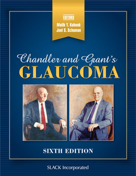
Chandler and Grant’s Glaucoma—one of the field’s seminal texts on the medical practice and surgical management of glaucoma, now in its Sixth Edition—includes the latest updates in an area that is currently experiencing a surge of innovation.
Edited by Drs. Malik Y. Kahook and Joel S. Schuman and with writings from the late Dr. David L. Epstein and more than 80 contributors, Chandler and Grant’s Glaucoma, Sixth Edition brings together the experience of world-class glaucoma experts who belong to a long line of surgeons trained using the teachings of the original authors of this classic work.
Each chapter has been meticulously edited and updated from the previous edition, while maintaining the well-established historical teachings of Drs. Paul A. Chandler and W. Morton Grant. New chapters on medical therapy as well as thorough updates on novel and minimally invasive approaches for the surgical management of glaucoma have been added.
New topics and features in the Sixth Edition include:
- Advances in imaging of the optic nerve and retina
- Rho-associated protein kinase inhibitors
- Glaucoma and cerebrospinal fluid pressure
- The FDA’s role in the development of new diagnostic and surgical devices for patients with glaucoma
More Information
Contents
DedicationAbout the Editors
Contributing Authors
In Memoriam
A Look Back to the Preface to the Fifth Edition
Preface to the Sixth Edition
Introduction
Section I The Basics
Chapter 1 Introduction
Chapter 2 Anatomy
Peripapillary Atrophy
Chapter 3 Practical Aqueous Humor Dynamics
An Interesting Question
Testing of New Glaucoma Drugs
Chapter 4 The Patient’s History: Symptoms of Glaucoma
Steroid-Induced Elevation of Intraocular Pressure
Chapter 5 Examination of the Eye
The Rule of 5%
Chapter 6 Tonometry and Tonography
Chapter 7 The Angle of the Anterior Chamber
Gonioscopy During Operation
Chapter 8 Examination of the Optic Nerve
Clinical Assessment of the Nerve Fiber Layer
Chapter 9 Imaging the Optic Nerve Head, Peripapillary, and Macula Regions in Glaucoma
Chapter 10 Imaging Devices for Angle Assessment
Chapter 11 Visual Fields and Their Relationship to the Optic Nerve
Section II Medications Used in Glaucoma Therapy
Chapter 12 Adrenergic Agents: Blockers and Agonists
Evaluating Clinical Trials
Timolol and Albino Rabbits
Laboratory Glaucoma Models
Statistical and Clinical Significance
Mechanisms of Epinephrine-Induced Cystoid Macular Edema
Chapter 13 The Miotics
Acetylcholinesterase
Drug Therapy Compliance
Limited Duration of Activity of Phospholine Iodide
Chapter 14 Carbonic Anhydrase Inhibitors: Systemic Use
Chapter 15 Topical Carbonic Anhydrase Inhibitors
Chapter 16 Prostaglandin Analogs
Prostaglandin Analogs and Sustained-Release Drug Delivery Platforms
Chapter 17 Rho-Associated Protein Kinase Inhibitors and Primary Open-Angle Glaucoma
The Discovery of ROCK Inhibitors as a Treatment for POAG
Chapter 18 Management of Highly Elevated Intraocular Pressure
Section III Common Open-Angle Glaucomas
Chapter 19 Primary Open-Angle Glaucoma
Primary Open-Angle Glaucoma and Myopia
Common Mistakes in Glaucoma Management
Trabecular Glaucoma Versus Optic Nerve Glaucoma
Tonography
Chapter 20 Normal-Tension Glaucoma
Normal-Tension Glaucoma and the General Ophthalmologist
Ocular Effects of Calcium-Channel Blockers: Past Promise
Chapter 21 Pseudoexfoliation Syndrome and Open-Angle Glaucoma
Tonography in Pseudoexfoliation
Chapter 22 Pigment Dispersion and Pigmentary Glaucoma
Tonography
Section IV Angle-Closure Glaucomas
Chapter 23 Principles of Primary Angle-Closure Glaucoma
Iris Retraction Syndrome
Pseudo-Unilateral Angle Closure
Chapter 24 Acute Angle-Closure Glaucoma: Diagnosis and Treatment
Anterior Chamber Deepening With Mechanical Breaking of Peripheral Anterior Synechiae With an Iris Spatula (Goniosynechialysis)
Chapter 25 Subacute (and Chronic) Angle-Closure Glaucoma
Chapter 26 Angle-Closure Glaucoma: Evaluation and Treatment After Iridotomy
Chapter 27 Plateau Iris
Chapter 28 The Use of Special Tests in Narrow-Angled Eyes
Goniolens Characteristics
Section V Secondary Angle-Closure Glaucomas
Chapter 29 Principles of Secondary Angle-Closure Glaucomas
Chapter 30 The Malignant Glaucoma Syndromes
The History of Mydriatic-Cycloplegic Therapy
The Chandler Operation (1965)
Chapter 31 Nanophthalmos: Diagnosis and Treatment
Chapter 32 Neovascular Glaucoma
Chapter 33 Iridocorneal Endothelial Syndrome
Chapter 34 Glaucoma After Vitreoretinal Procedures
Intraocular Gas and Altitude
Chapter 35 Angle-Closure Glaucoma Due to Multiple Cysts of the Iris and Ciliary Body
Chapter 36 Angle-Closure Glaucoma Secondary to Occlusion of the Central Retinal Vein
Chapter 37 Angle-Closure Glaucoma Secondary to Acute Myopia
Acute Bilateral Transitory Myopia Associated With Open-Angle Glaucoma
Chapter 38 Glaucoma After Penetrating Keratoplasty
Section VI Combined Mechanisms
Chapter 39 Combined Open-Angle and Angle-Closure Glaucoma
Chapter 40 Glaucoma in the Pseudophakic and Aphakic Eye
Glaucoma Surgery in Pseudophakia and Aphakia: A Historical Perspective
Chapter 41 Characteristically Unilateral Glaucomas: Differential Diagnosis
Chapter 42 Glaucoma Secondary to Intraocular Tumors
Section VII Secondary Open-Angle Glaucomas
Chapter 43 Glaucoma Due to Intraocular Inflammation
Chapter 44 Glaucoma Due to Trauma
Secondary Glaucoma in Black Patients: The Importance of Sickling:
Iris Retraction Syndrome
Chapter 45 Corticosteroid Glaucoma
Chapter 46 Hemolytic or Ghost-Cell Glaucoma
Chapter 47 Glaucoma Associated With Extraocular Venous Congestion (Increased Episcleral Venous Pressure)
Aqueous Humor Dynamics
Chapter 48 Lens-Induced Glaucoma
Understanding Lens-Induced Glaucomas
Completely Dislocated Hypermature Cataract and Glaucoma
Chapter 49 Amyloidosis and Open-Angle Glaucoma
Chapter 50 Glaucoma in the Phakomatoses
Chapter 51 Juvenile Open-Angle Glaucoma
Section VIII Laser Methods in Glaucoma
Chapter 52 Glaucoma Laser Surgery
Chapter 53 Laser Trabeculoplasty
Chapter 54 Laser Trabeculoplasty: How Does It Work?
How I Do Laser Trabeculopasty
Chapter 55 Post-Laser Elevation of Intraocular Pressure
Chapter 56 Laser Peripheral Iridotomy
The Choice of Iridotomy Lens
Technique of Laser Iridotomy
One Technique for Laser Peripheral Iridotomy
Which Laser Should I Use?
Chapter 57 Cyclodestruction
Chapter 58 Laser Peripheral Iridoplasty
Practical Considerations for Argon Laser Peripheral Iridoplasty
Personal Technique
Section IX Glaucoma Surgery
Chapter 59 What to Say to Patients With Glaucoma Prior to Filtration Surgery
Chapter 60 Filtering Surgery in the Management of Glaucoma
Antiproliferative Therapy for Filtration Surgery
Externalized Releasable Sutures in Filtering Surgery
How to Handle Mitomycin C
Chapter 61 Postoperative Management Following Filtration Surgery
Performing a Choroidal Tap
Treatment of Hypotonous Maculopathy
Chapter 62 The Management of Coexisting Cataract and Advanced Glaucoma
The Management of Coexisting Cataract and Mild to Moderate Glaucoma
Chapter 63 Aqueous Shunting Procedures
Chapter 64 Cyclodialysis
Mystery Diagnosis
Chapter 65 Surgical Peripheral Iridectomy
Chapter 66 Schlemm’s Canal Surgery for Glaucoma Management
Goniotomy for Treatment of Glaucoma in Adults
Chapter 67 Suprachoroidal Approach to Glaucoma Surgery
Chapter 68 Treatment of Occludable Angles and Angle Closure With Cataract Extraction
Section X Diagnosis and Treatment of Glaucoma in Children
Chapter 69 Pediatric Glaucoma
Chapter 70 Unusual Pediatric Glaucomas
Section XI Special Considerations
Chapter 71 The Role of the Cornea in Managing Glaucoma
Chapter 72 Twenty-Four–Hour Intraocular Pressure Monitoring in Glaucoma
Chapter 73 The Role of Ocular Perfusion Pressure in the Pathogenesis of Glaucoma
Topical Medications and Ocular Perfusion Pressure
Chapter 74 Glaucoma and Cerebrospinal Fluid Pressure
Chapter 75 Neuroprotection in Glaucoma
Chapter 76 Adherence to Glaucoma Medical Therapy
What Do We Actually Do to Ensure Adherence?
Chapter 77 The FDA’s Role in the Regulation of New Diagnostic and Surgical Devices
Financial Disclosures
Index
About the Editors
Malik Y. Kahook, MD is Professor of Ophthalmology and The Slater Family Endowed Chair in Ophthalmology at the University of Colorado School of Medicine. He is Vice Chair of Translational Research and serves as chief of the glaucoma service and co-director of the glaucoma fellowship at the UCHealth Sue Anschutz-Rodgers Eye Center.Dr. Kahook has authored more than 400 peer-reviewed manuscripts, abstracts, and book chapters and is editor of Essentials of Glaucoma Surgery; MIGS: Advances in Glaucoma Surgery; and the seminal textbook of glaucoma Chandler and Grant’s Glaucoma. He is Editor-in-Chief of the open access wiki-based glaucoma educational platform Kahook’s Essentials of Glaucoma Therapy (www.KEOGT.com). Dr. Kahook has received funding from the National Eye Institute, Foundations and Industry over the past 14 years. He was awarded an American Glaucoma Society Clinician-Scientist Fellowship Award in 2007 as well as the American Glaucoma Society Compliance Grant in 2006 and was named New Inventor of the Year for the University of Colorado in 2009 and Inventor of the Year for 2010. He received the American Glaucoma Society Innovator Award (2020), the American Academy of Ophthalmology Achievement Award in 2011, the American Academy of Ophthalmology Senior Achievement Award in 2017, the American Academy of Ophthalmology Secretariat Award (2014), the Ludwig Von Sallmann Clinician-Scientist Award (ARVO) in 2013 and was ranked second on the 40 under 40 Ophthalmology Power List (2015). Dr. Kahook has served on several editorial boards including the American Journal of Ophthalmology and International Glaucoma Review. He is an active volunteer with Orbis and has been a consultant to the US Food and Drug Administration’s Ophthalmic Device Division since 2008.
Dr. Kahook has been active in both basic and clinical research. He and his colleagues were among the first to report the use of anti–vascular endothelial growth factor (VEGF) agents for treating neovascular glaucoma, as well as the first to report the use of these agents to modulate blebs after filtration surgery. Dr. Kahook and colleagues explored the effects of anti-VEGF agents on the trabecular meshwork and published a series of papers that explored the potential for intraocular pressure spikes from contaminants in compounded anti-VEGF syringes. His work exploring silicone microbubbles in repackaged bevacizumab shed light on several compounding pharmacy practices, including shipping techniques and freeze thaw cycles that resulted in recommendations to decrease the chance of these potentially harmful contaminants from reaching the patient. Dr. Kahook is widely published in areas ranging from the effects of medication preservatives on the ocular surface to novel imaging techniques with femtosecond lasers, as well as 24-hour intraocular pressure fluctuations and exploration of adherence to medical therapy. His first report with Dr. Robert Noecker on the use of fibrin glue for glaucoma drainage device surgery has been adopted by surgeons globally.
Dr. Kahook’s translational research accomplishments have focused on multiple unmet needs, including advanced cataract surgery devices and implants, novel glaucoma therapies, and advanced imaging techniques. He has filed for more than 120 patents with more than 40 patents granted to date. Several of Dr. Kahook’s patents have been licensed by companies including Johnson & Johnson Vision, New World Medical, ShapeTech, Alcon, ClarVista Medical, and SpyGlass Ophthalmics for development and commercialization. Six of his devices are currently in human trials or commercialized for clinical use. ClarVista Medical, acquired by Alcon in 2017, developed an advanced intraocular lens technology platform invented by Dr. Kahook. He is also inventor of the Kahook Dual Blade, which is marketed globally by New World Medical. Dr. Kahook is also inventor of the VERUS Capsulorhexis Device, commercialized by MileHigh Ophthalmics, and the ShapeTech shape memory polymer intraocular lens material, which is licensed by Johnson & Johnson Vision. His inventions have raised more than $100 million for development and commercialization since 2008 and have been used to treat more than 100 thousand patients globally since 2012.
After graduating from Northeastern Ohio Universities College of Medicine, Dr. Kahook completed his residency training at the University of Colorado, Rocky Mountain Lions Eye Institute in Denver, Colorado, where he was named Chief Resident. He then went on to complete a fellowship in glaucoma with Joel S. Schuman and Robert J. Noecker at the University of Pittsburgh Medical Center in Pittsburgh, Pennsylvania.
Joel S. Schuman, MD, FACS is the Elaine Langone Professor and Vice Chair for Research in the Department of Ophthalmology and Professor of Neuroscience & Physiology at NYU Langone Health, NYU Grossman School of Medicine. He is Professor of Biomedical Engineering and Electrical & Computer Engineering at NYU Tandon School of Engineering and Professor of Neural Science in the Center for Neural Science at NYU College of Arts & Sciences. He chaired the ophthalmology department at NYU Langone Health, NYU Grossman School of Medicine (2016-2020). Prior to arriving at NYU in 2016, he was the Eye and Ear Foundation Professor and Chairman of Ophthalmology (2003-2016), the Eye and Ear Institute, University of Pittsburgh School of Medicine, and Director of the University of Pittsburgh Medical Center (UPMC) Eye Center, Professor of Bioengineering at the Swanson School of Engineering, University of Pittsburgh, and Founder of the Louis J. Fox Center for Vision Restoration of UPMC and the University of Pittsburgh. He was a member of the McGowan Institute for Regenerative Medicine and the Center for the Neural Basis of Cognition, Carnegie Mellon University, and University of Pittsburgh. Dr. Schuman is a native of Roslyn, New York; he graduated from Columbia University (AB, 1980) and Mt. Sinai School of Medicine (MD, 1984). Following his internship at New York’s Beth Israel Medical Center (1985), he completed residency training at Medical College of Virginia (1988) and glaucoma fellowship at Massachusetts Eye and Ear Infirmary (clinical 1989; research 1990), where he was a Heed Fellow. After just over a year on the Harvard faculty, he moved to New England Medical Center, Tufts University, to co-found the New England Eye Center in 1991, where he was Residency Director (1991-1999) and Glaucoma and Cataract Service Chief (1991-2003). In 1998 he became Professor of Ophthalmology, and Vice Chair in 2001.
Dr. Schuman and his colleagues were first to identify a molecular marker for human glaucoma, published in Nature Medicine in 2001. Continuously funded by the National Eye Institute as a principal investigator since 1995, he is an inventor of optical coherence tomography, used worldwide for ocular diagnostics. Dr. Schuman has published more than 400 peer-reviewed scientific journal articles, has authored or edited 8 books, and has contributed more than 80 book chapters. Dr. Schuman is a founding member of the Association for Research in Vision and Ophthalmology (ARVO) Multidisciplinary Ophthalmic Imaging cross-sectional group, served on the program committee from its founding, and chaired the MOI program committee 2007-2013. He is also a founder and co-chair of ARVO Imaging (formerly ARVO/isie, the International Society for Imaging in the Eye, inaugurated 2002). Dr. Schuman was co-chair of the International Glaucoma Symposium 1998-2007, the world’s largest meeting devoted to glaucoma, which merged with the World Glaucoma Congress in 2007, for which he was Program co-chair 2007-2011. He chaired the Hawaiian Eye meeting glaucoma section 1993-2019.
In 2002 he received the Alcon Research Institute Award and the Lewis Rudin Glaucoma Prize, in 2003 the Senior Achievement Award from the American Academy of Ophthalmology, in 2004 he was elected into the American Society for Clinical Investigation, in 2006 he received the ARVO Translational Research Award, and in 2012 the Carnegie Science Center Award, as well as sharing the Champalimaud Award (a 1 million Euro cash prize). He was elected to the American Ophthalmological Society in 2008. In 2011 Dr. Schuman was the Clinician-Scientist Lecturer of the American Glaucoma Society. In 2012 he received the Carnegie Science Center’s Award in Life Sciences. In 2013 he gave the Robert N. Shaffer Lecture at the American Academy of Ophthalmology Annual Meeting, and received the Academy’s Lifetime Achievement Award. In 2014 he became a Gold Fellow of ARVO and he received a Special Recognition Award from the American Academy of Ophthalmology in 2015. He was elected to the American Association of Physicians and also received the Fight for Sight Physician/Scientist Award in 2016. In 2017 he received the Leslie Dana Gold Medal. In 2018 Dr. Schuman was the American Glaucoma Society Lecturer and received the Fight for Sight Alumni Achievement Award. In 2019 he was given the BrightFocus Scientific Impact Award. He is named in Who’s Who in America, Who’s Who in Medical Sciences Education, America’s Top Doctors, and Best Doctors in America.

